
Blog article - From planning to perfection: the journey of a facial contouring implant
Introduction
Facial contouring surgery requires precise knowledge and advanced tools to meet both functional and aesthetic goals. The rise of patient-specific implants (PSIs) has revolutionized this field, enabling surgeons to craft solutions tailored to each individual. From virtual planning to post-surgical outcomes, PSIs like MyBone® offer a seamless journey that ensures stable results and patient satisfaction. This article delves into the journey of creating and implementing a MyBone® facial contouring PSI, showcasing how innovation transforms surgery at every stage.
Step 1: Virtual surgical planning
The process begins with virtual surgical planning (VSP), a cornerstone of modern maxillofacial surgery. Using high-resolution 3D imaging techniques, such as computed tomography (CT) scans, surgeons can visualize the patient’s anatomy with unparalleled clarity (Parthasarathy, 2014) and provide the engineering team with a very clear and specific image. Subsequently, advanced software tools allow for precise mapping of asymmetries, enabling accurate planning and design of the implant.
What sets Cerhum apart is our collaborative approach—we don’t just create patient-specific implants; we co-design them with surgeons. By working closely with each practitioner, we ensure that the implant is not only tailored to the patient’s anatomy but also optimized for the surgeon’s specific technique and surgical approach. This synergy results in a seamlessly integrated implant solution, improving surgical efficiency, predictability, and patient outcomes.
Step 2: Manufacturing the implant
Once the planning and design phase is complete, the next step is manufacturing the implant. Cerhum’s proprietary 3D printing technology, uniquely developed in-house, pushes the boundaries of bioceramic precision, achieving a level of accuracy and detail previously unattainable in this field. By leveraging this innovation with bioceramic materials like hydroxyapatite—a trusted material in surgery for years—MyBone® takes its potential to the next level, offering unprecedented precision, integration, and reliability in patient-specific implant design, making it a leading choice for facial contouring (Systermans et al., 2024).
Key features of MyBone® implants
- Mimics bone: MyBone is made from hydroxyapatite, the main component of natural bone, which reduces risks of infection or rejection..
- Superior porosity: with up to 70% porosity, the implant’s pore size and geometry are carefully engineered to promote vascularization and osseointegration, ensuring faster healing and long-term stability.
- Tailored aesthetics: each implant is designed for precise symmetry and soft tissue balance, addressing the specific needs of facial contouring (Verbist et al., 2024).
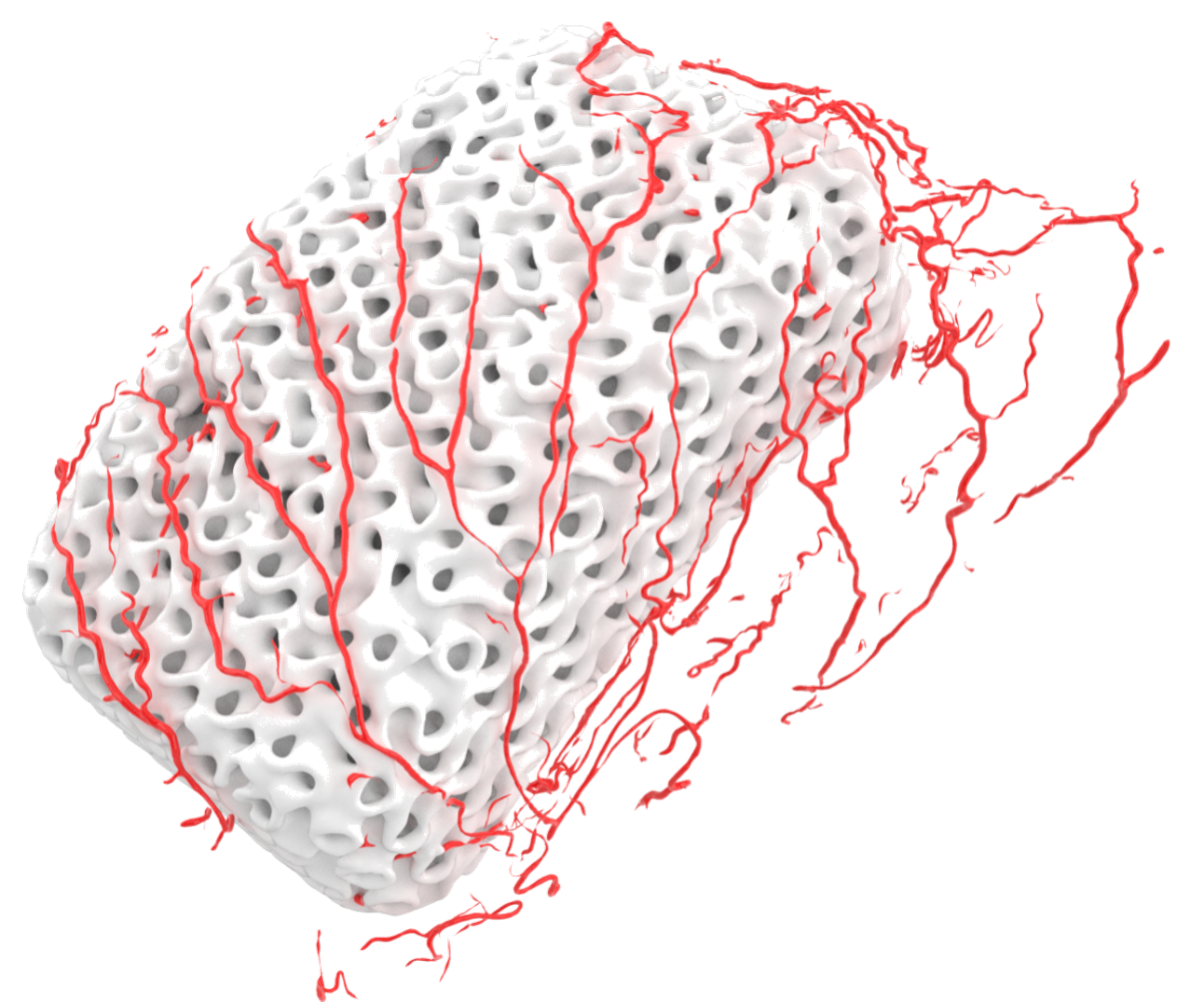
MyBone Custom: unique combination of an osseoconductive material with an optimized porosity promoting vascularization and osseointegration
Step 3: Surgical implementation
Intraoperative precision is crucial to the success of facial contouring interventions. The implant’s patient specificity, resulting from the previous phase, simplifies and shortens the surgery process for surgeons, reducing the need for adjustments during the procedure. Additionally, the drillable and adaptable nature of the material allows for real-time refinements if needed, further enhancing outcomes (Modgill et al., 2023).
Step 4: Recovery and results
Postoperative recovery is often smoother with MyBone® implants. Imaging studies conducted after surgery consistently show proper integration of the implant with the surrounding bone, as highlighted in a series of case studies focused on MyBone®. Furthermore, patients experience fewer complications, faster healing times, and predictable aesthetic results, with lasting symmetry and natural-looking features, catered to their individual needs.
A case in point: MyBone® in action
Dr. Beckers utilized MyBone® Custom in a relevant case involving a 33-year-old female patient presenting with pronounced medial facial hypoplasia, requiring zygomatic augmentation. Rather than performing a Le Fort III osteotomy—a complex procedure with significant morbidity and extended recovery (Schlieder & Markiewicz, 2022), the surgeon opted for a patient-specific MyBone® hydroxyapatite implant (HA-PSI) designed for precise augmentation.
The implant was designed for optimal zygomatic projection and structural support, featuring a single screw fixation (Ø 2.0 mm) for enhanced stability. The careful preoperative planning allowed for precise implant placement, ensuring both aesthetic enhancement and long-term functional integrity.
During the procedure, the patient-specific nature of MyBone® minimized intraoperative adjustments, streamlining the surgery. The biocompatibility of the hydroxyapatite material enabled seamless integration with the patient’s existing bone, eliminating concerns associated with traditional materials like PEEK or titanium.
The results showcased enhanced facial symmetry and aesthetic balance, with successful integration and no reported complications. Avoiding a more invasive procedure significantly improved recovery, and the patient expressed high satisfaction with the natural, refined outcome.
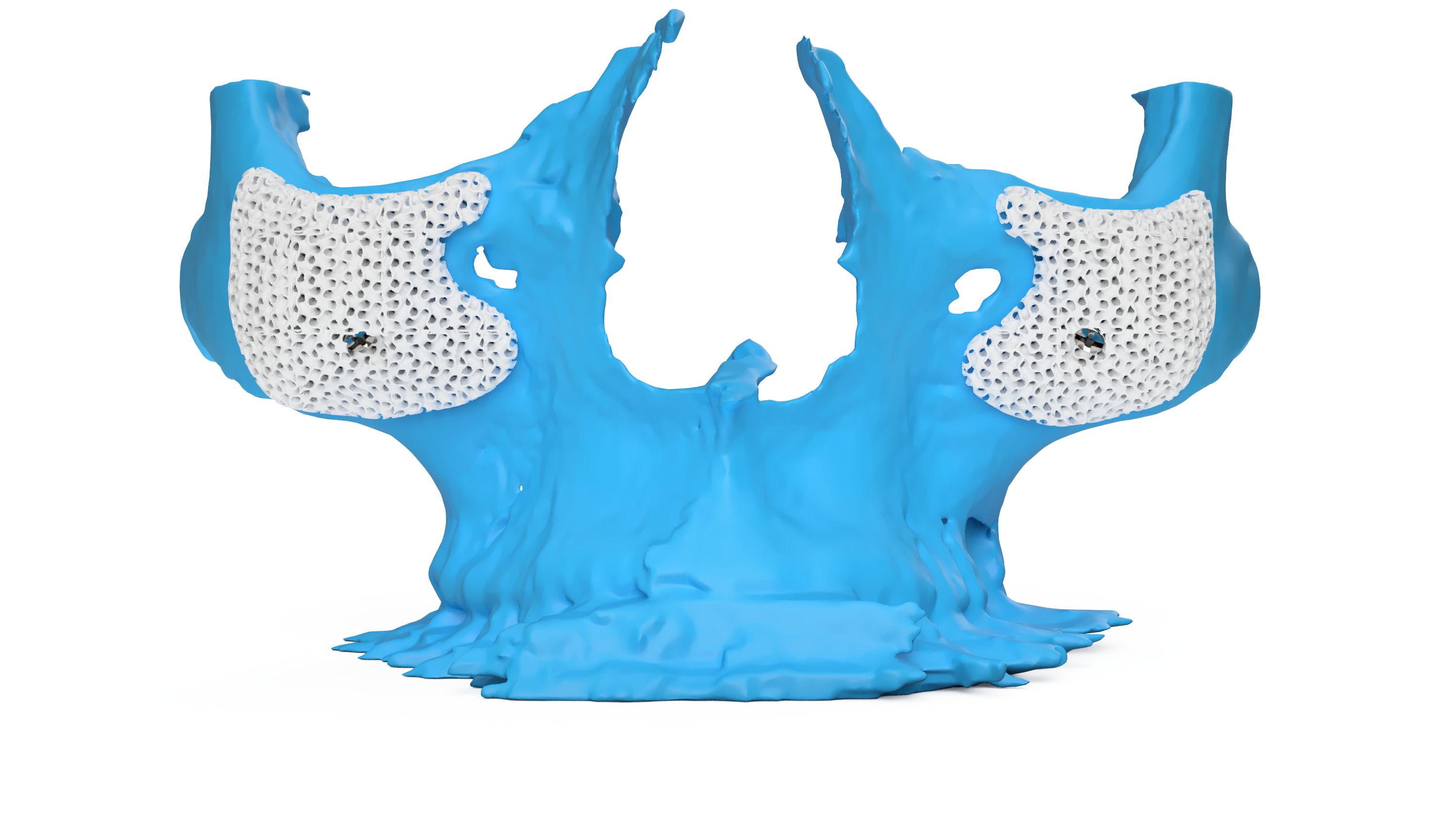
Rendering of the virtual surgical planning.
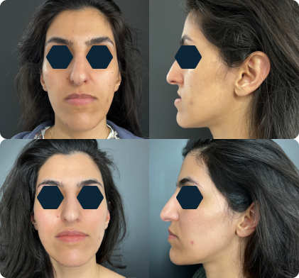
Surgical outcome - Before and after comparison.
Conclusion
The journey of a facial contouring implant, from planning to perfection, exemplifies the transformative power of innovation in maxillofacial surgery. MyBone® stands at the forefront of this evolution, combining advanced technology, superior materials, and patient-specific designs to deliver exceptional results. As surgeons increasingly adopt PSIs, the future of facial contouring promises even greater precision, efficiency, and patient satisfaction.
To find out more about how MyBone® can support your facial contouring procedures, get in touch with us.
References
● Parthasarathy, Jayanthi. 3D modeling, custom implants and its future perspectives in craniofacial surgery. Annals of Maxillofacial Surgery 4(1):p 9-18, Jan–Jun 2014. .doi:10.4103/2231-0746.133065
● Parthasarathy, J. (2014). 3D Modeling, Custom Implants and Its Future Perspectives in Craniofacial Surgery. Annals of Maxillofacial Surgery, 4(1), 9-18.
● Systermans, S., et al. (2024). An Innovative 3D Hydroxyapatite Patient-Specific Implant for Maxillofacial Bone Reconstruction: A Case Series of 13 Patients. Journal of Cranio-Maxillofacial Surgery, 52(420-431). https://doi.org/10.1016/j.jcms.2024.02.026
● Verbist, M., et al. (2024). Reconstruction of Craniomaxillofacial Bone Defects with 3D-Printed Bioceramic Implants: Scoping Review and Clinical Case Series. Journal of Clinical Medicine, 13(2805). https://doi.org/10.3390/jcm13102805


